


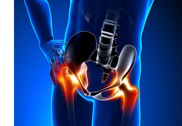
Arthrosis of the hip joint (Coxartrosis)- This is a chronic degenerative joint disease, which leads to the deformation of the bone tissue.With Coksartrosis, all components of the joint are involved in the pathological process: joint cartilage, bone structures adjacent to cartilage, synovial shell, ligaments, capsule and adjacent muscles.In case of illness, the joint cartilage is destroyed, micro-inghemies of bones and osteophytes appear (bone outcrops) and an inflammation of the muscle-legal system of the hip joint occurs.
In the world, every fifth person complains about joint problems with the joints.This can be both the pain or restriction of the movement in the joints, and a combination of these symptoms.Every second outpatient vision falls on patients with bone-muscular disorders, while 66 % of cases have people under the age of 65.According to the latest epidemiological research, the prevalence of arthrosis of the knee and hip joints between the adult population is 13 %.
Risk factors for the development of coxarthrosis:
- Genetic predisposition.A common cause of coksartrosis of the hip joints is the congenital or acquired mutation of the type of type II prollagen.
- Elderly.The probable cause of the prevalence of arthrosis in old age is a discrepancy between the harmful effect on the joint cartilage of the external environment and its ability to restore.
- Flooring.Women suffer from osteoarthritis more often than men.This is due to the effects of the influence of female sex hormones of estrogen on the metabolism of bone mineral.However, the influence of the floor is ambiguous - according to some authors, unlike the damage to other joints, there are no differences based on Coksartrosis: in men, the arthrosis of the hip joint is often found as in women.
- Excess body weight.The relationship is demonstrated between the excess body mass and the occurrence of arthrosis.The excess adhesive tissue increases the harmful load on the cartilage.In addition, adipose tissue produces pro -inflammatory enzymes that damage the cartilage tissue.
- Frequent development of bones and joints.In accordance with studies, 80 % of coxartrosis, which occurs without apparent reason, is associated with previously not diagnosed defects in the development of the hip joint - dysplasia and subluxation.
- Heavy physical work.An excess load on the hip joints with certain types of physical work can cause damage to the joints and the formation of arthrosis.At risk are agricultural workers, diggers and people with similar work specialties.
- Injuries.The risk of developing coxartrosis increases after an injury to the hip joint.Furthermore, it is an injured joint that both can be involved in the process.
- Professional gaming sport.Professional sport can cause the occurrence of coxarthrosis both due to the excessive load on the joints and for injuries.Potentially dangerous sports include heavy athletics, athletics leap, parachute sports.
- Bones and joint diseases- Rheumatoid arthritis, psoriatic arthritis, joint infections, avascular necrosis, gottosa arthritis, etc.
- Endocrine pathologies- hypothyroidism, hypoparatyroidism, acromegaly (compromised function of the front pituitary gland), diabetes, obesity.
If similar symptoms are detected, consult a doctor.Don't trundiare: it's dangerous for your health!
Symptoms of arthrosis of the hip joints
The main symptoms of coxartrosis include: pain, mobility restrictions and creaking in the joints, their deformation, the functional shortening of the lower limb and the periodic swelling in the joints.
Pain of various intensity.The pain in the joint is initially insignificant and rises for a short period.They seem or intensify during walking or with other physical efforts, for example during squats, inclinations and weight lifting.As the disease develops, pain intensifies and even a long rest does not bring relief.In addition, the pain occurs with prolonged immobility and the fixation of the joint in a position.
Patients complain of pains so called "initial" in the hip joints after sleeping, guiding in a car and other prolonged immobility.The "start" pain for coxartrosis does not last more than 30 minutes.Pain intensifies during hypothermia or in a stressful situation.They can be located in the glute or grove area, on the front or lateral surface of the thigh.With the spread of pain on the nerves of the lumbar plexus, it can be transmitted to the thighs distant from the center of the body or knee.Sometimes the pain applies to the lumbosacral column and coccyx.
Limitation of joint mobility.The movements in the hip joint with Coksartrosis are limited due to pain.At the same time, the rotation (turns both inside and outside) and bringing the lower limb (movement to the center of the body) are more often disturbed, but it can be limited (movement from the average axis of the body), as well as flexion and extension.The inability to carry out passive movements in the joint due to a pronounced pain syndrome causes a compensatory pelvic distortion.The patient's pace changes, the buttocks protrude back, the body differs forward when it transfers the weight to the damaged side.With bilateral damage in patients with coksartrosis, a patient of duck is formed ".
With Coxartrosis it occurs periodicallyswelling in the jointwhich can be invisible due to the muscle and the layer of fat.In addition, the disease is characteristicCrystal in the joints during the movement, their gradual deformation and the functional shortening of the lower limb.
Often, an articulation is influenced by the disease, so the process applies to others.But sometimes arthrosis affects several joints simultaneously and polyosostoarthritis occurs.Poliosteoartrosis is characteristic of the elderly or with a hereditary predisposition and concomitant diseases: diseases of bones, joints and endocrine disorders.
Pathogenesis of the arthrosis of the hip joints
In the pathogenesis of arthrosis of the hip joints, an important role is played by mechanical damage (injuries and microtrauma due to an increase in physical effort on the articulation) and genetic, hormonal and metabolic factors.Often it is not possible to find out which factor has influenced the development of the disease in a particular patient, but often the disease develops after tissue damage with mechanical injuries.
The damage to the tissue stimulates the division of the cells of the cartilage tissue (chondrocytes), while the production of cytokines pro -inflammatory, which are normally present in the cartilage only in small quantities increases.The cytokines launch the inflammatory process, for example, under the influence of the IL-1 pro-inflammatory cytokines, the enzymes are distinguished that they destroy the cartilage of the joint.In addition, under the influence of cytokines, the production of the tsog-2 enzyme and other substances that have a toxic effect on the increases of the cartilage.
The Sinovites also play an important role in the development of coxartrosis: inflammatory diseases of the synovial shell of joints or ligaments with the accumulation of liquid in the cavity.
A decrease in the elasticity and strength of the joint cartilage associated with metabolic disorders leads to a decrease in its resistance to mechanical stress.With Coksartrosis, all components of the joints are involved in the pathological process, including a sub -controlle bone.Due to the fact that the large joints of the lower ends represent great joints of the body, they experience a significant mechanical stress, due to which micro -valums occur in the plate and in the subcontracting cartilage.As a result of the microvelomas, the subcondral bone is compact, which leads to the regional growth of bone tissues.And this, in turn, stimulates the further degradation of the joint cartilage.
In some cases, the arthrosis of the hip joint is inherited.Hereditary arthrosis is presumably a polygraenic legacy - due to the action of many genes, each of which affects weakly.The cause of some diseases is a mutation in the genes that codify the macromolecules of the joint cartilage, which causes its breakages.The genes responsible for the division of chondrocytes can also suffer.In addition, metabolic disorders are inherited, such as the arthropathy of pyrophosphate - a disease in which calcium pyrophosphate crystals accumulate in the joint cartilage and in the synovial liquid.
Classification and phases of development of the arthrosis of the hip joints
Depending on the causes of the disease, coxartrosis is divided into two main forms: primary (idiopathic) and secondary (deriving from or due to other diseases).
Primary Coksartrosis:
- Localized (only hip reached):
- unilateral;
- bilateral.
- Generalized (Poliosteoartrosi) with an injury of at least three joint groups (for example, hip, knee and small joints of brushes or feet).
Secondary arthrosis:
- Post -traumatic:
- acute - as a consequence of the acute injury;
- Chronic - Due to the classes of some sports or as a result of professional activity.
- Metabolic diseases (occonance, bloodocromatosis, Wilson's disease, Gaucher's disease).
- Congenital pathologies and development defects (congenital dysplasia of the hip joint, pearl disease, sliding of the epiphae of the femur, hypermability syndrome, shortening of the lower limb, scoliosis, bone dysplasia).
- Endocrine pathologies (acromegaly, hypothyroidism, diabetes mellitus, hyperparathyroidism, obesity).
- Football salts (arthropathy of pyrophosphate, Calcifying tendonitis).
- Bone diseases and joints (rheumatoid arthritis, psoriatic arthritis, pedestic disease, avascular necrosis, infections).
According to clinical manifestations, the following forms of coxartrosis are distinguished:
- Little symptomic.
- Manifesto, manifested by bright clinical symptoms:
- quickly progressive, in which the symptoms develop in the first four years from the beginning of the disease;
- Slowly progressive - clinically significant symptoms appear after five years of the course of the disease.
In accordance with the image of X -rays, it is possible to identify two types of arthrosis of the hip joints:
- Hypertrophic - With signs of increasing reparative response (the lesions are replaced by a new tissue, for example, osteophytes appear);
- Atrophic (decrease in the volume of the fabric).
The phases of the disease can be determined radiologically and clinically.To determine the radiological stage of arthrosis of the hip joint, the classification of Kellgren and Lawrence is often used (1957).
Highth phase in the radiological classification
| Stage | Signs |
|---|---|
| 0 | There are no signs of arthrosis in the X -ray images |
| 1 | The joint gap is not changed, single osteophytes are displayed |
| 2 | The joint gap is not changed, significant regional osteophytes are displayed |
| 3 | The height of the joint gap is moderately reduced, significant regional osteophytes are displayed |
| 4 | The height of the joint gap is significantly reduced, significant regional osteophytes are displayed and subcontracting osteosclerosis (comparison of the bone tissue under the lower surface of the cartilage with the structure of the cartilage) |
To determine the clinical stage of the disease, classification is used (1961), which uses both clinical signs and visualization criteria.
Clinical phase of arthrosis
| Stage | Signs |
|---|---|
| 0 | The joint gap is unequivocal and irregularly reduced, the edges of the joint cracks are slightly pointed (initial osteophytes), there is a slight restriction of the movements |
| 1 | The joint gap is significantly restricted (50-60 %), significant osteophytes, sub-controlle osteochosclerosis and cystic lighting in bone epiphytes;The clinic is predominated by the restriction of mobility in the joints, a rough creaking during movements, insignificant or moderate muscle atrophy |
| 2 | deformation, rigidity of the joint;The joint gap is reduced by over 60-70 % of the rule or completely absent, extensive osteophytes, sub-compliant cysts, joint "mice" are displayed bone, cartilage or mixed pathological formations located in the joint cavity |
Complications of the arthrosis of the hip joints
With coxartrosis, all complications are associated with pathological changes in the joints.
The course of Coksartrosis can be complicated by local inflammatory processes:
- Bursitis: inflammation of synovial bags in the joints;
- Tenting - inflammation of the internal shell of the muscle tendon vagina;
- Tunnel syndrome of the pinchi of the nerve due to the formation of large osteophytes or with joint deformation.
With the progression of coxartrosis and its transition to the clinical phases II and III, the pain limits the mobility of the joint and over time the joint ankylosis occurs (fibrous, bone or cartilage), accompanied by its complete immobility.
A significant joint deformation can lead toFractures or aseptic necrosis of the bones.For Coksartrosis, the aseptic necrosis of the femoral head is the most formidable complication.
With pronounced coks arthrosis, it can occurSubluxation and dislocation of the jointAs well as the penetration of the femoral head in the pelvic cavity.The dislocations and subluxation of the hip joint lead to pain (initially acute, therefore opaque and painful), intensifying during the walk and other physical efforts, as well as the deformation of the joint, to the lame and sometimes to shorten the limb concerned.
Despite the lack of systemic manifestations of arthrosis itself, in modern clinical practice, greater attention is paid to associated diseases.These are such pathological conditions that exist or arise against the background of the current disease.In relation to the inflammatory reactions deriving during arthrosis, the formation of atherosclerotic plates on the internal walls of the vessels has improved, which increases the riskCardiovascular diseases.A decrease in physical activity due to the pain and restriction of joint mobility leads toObesity, depression and deterioration of the quality of life.With prolonged use of non -pounded anti -inflammatory drugs,The upper gastrointestinal sections are interested,And alsoThe risk of cardiovascular pathologies and kidney disease increases.
Diagnosis of arthrosis of the hip joints
The diagnosis of "coksartrosis" is made on the basis of clinical manifestations and radiological examination.There are no characteristic laboratory signs for the diagnosis of arthrosis.
Among the clinical manifestationsThe main for the diagnosis of arthrosis of the hip joint is pain and its character.The pain for the arthrosis of the hip joint occurs and grows gradually for several years (sometimes several months with a quickly progressive form).Pain occurs or improves during physical effort or in the erect position.If the patient begins to feel pain alone, then inflammation (synovitis).The declaration is noted up to 30 minutes in the morning and with prolonged immobility.
The limitation of joint mobility is gradually increasing, this applies to both active and passive movements.With the development of the disease, the joints are deformed and a functional shortening of the length of the limbs can occur.
In an examination of physicinThere is a limitation of joint mobility, their deformation, the shortening of the limbs, the pain in the palpation of the joint and a large rotation of the femur, muscle atrophy.
Laboratory methodsFor the diagnosis of arthrosis of the hip joints are not necessary.However, they can be used for the differential diagnosis of coxartrosis with arthritis (rheumatoid and chronic), since with arthrosis there are no inflammatory changes in the examination of the overall blood and in the rheumatoid factor and the levels of uric acid have not increased.In addition, using laboratory tests, contraindications are revealed for pharmacological treatment methods.
Instrumental methodsFor the diagnosis of arthrosis of the hip joints:
- Radiography- This is the main method to diagnose the arthrosis of the hip joints.The X -ray determines the characteristic changes of Coksartrosis: narrowing of the joint gap, osteophyte, erosion and cartilage ulceration, subcontracting cysts and osteosclerosis.The X -Raist exam is a classic method for diagnosis of coxartrosis and radiological signs are the basis of the classification of coxartrosis.However, at the moment, other methods of visualization of the joint are increasingly used, such as ultrasound and magnetic resonance imaging.
- Ectrasuund examination (ultrasound) -The advantage of ultrasound is in the absence of a radial load on the body.
- Magnetic resonance imaging tomography (MRI)- Compared to other methods, it allows you to view the joint damage more clearly.
- Arthroscopy-It allows you to identify the damage to the joint cartilage: from the condar areas (softening of the joint cartilage) with a diameter of less than 10 mm to deep cracks that penetrate the subcontracting bone and the formation of deep ulcers.Surface and medium cracks can also be displayed and the erosion of the surface.
The identification of Coksartrosis usually does not represent special difficulties, but when evaluating a specific clinical situation, it is necessary to remember the possible secondary origin of the arthrosis of the hip joints (as complications of other diseases, for example, with endocrine disorders).
Treatment of arthrosis of the hip joints
The treatment of arthrosis of the hip joints can be both conservative (drug and not united) and operational.Conservative treatment is used in 1-2 stadiums of the disease, surgical stadiums.Surgical treatment can be recommended in 2 phases with persistent pain and lack of reaction to conservative therapy.
The objectives of conservative therapy:
- Improve the quality of life: reduce pain and increase joint mobility;
- Stop or slow down the development of the disease.
Non -Drug treatment methods include:
- Download the hip joint (decrease in body weight, the creation of additional support and the transfer of part of the body weight to the rod or crutches);
- physical education of physiotherapy;
- Physiotherapy treatment methods.
Coxartrosis treatment begins with non -pharmacological methods, an important role is assigned to physiotherapy exercises.With severe pain, the patient should use support.With a pronounced disease and the presence of contraindications to endoprothetics, the support must be used for life.
Cuxaartrosis medicinal therapyIncludes drugs that reduce the symptoms of the disease.These are analgesics, as well as drugs of the group of non -pound anti -inflammatory drugs (NSAIDs).Fans are divided into non -electoral and selective.
Analgesics and fans for the arthrosis of the hip joint are used for a short period to relieve pain and inflammation.Currently, there is no advantage proven by an anti -inflammatory agent non sterileidal compared to another, therefore the choice of a particular drug depends on the side effects and a specific clinical situation caused by it.
It should be remembered that fans have a series of side effects.When you take them, the mucous membrane of the mucous membrane of the stomach and the duodenum is affected, due to which ulcers and bleeding are possible.Numerous fans have a toxic effect on the liver and kidneys.In addition, fans interrupt platelet aggregation and, consequently, the patient is interrupted by thrombosis and there is a tendency to bleeding.Fans with prolonged use suppress the processes of the hematopoiesis and can cause aplastic and agranulocytosis anemia.The reception of selective fans causes significantly less complications.
The ointments and gels used locally cause less side effects than oral products.For the treatment of arthrosis, drugs with heating and reduction of pain are used.They may contain terrible, menthol, nicotinic acid foreign, salicylated, bee poison.In addition, fans have a good effect.
In the absence of the effect of analgesics and fans or if it is impossible to choose the optimal dose of the drug, the pain relievers of the central action can be prescribed in the short term.
In case of inflammation, intra -articular corticosteroid administration is used.Corticosteroids are used no more than 2-3 times a year, since more frequent use can lead to the degeneration of the cartilage.
The drugs with action slowly weaken the symptoms of the disease include chondroprotectors, inappropriate compounds of avocado or soybeans, hyaluronic acid.These drugs are included in the recommendations of the European Antirematic League for the treatment of arthrosis of the hip joints.Preparations reduce pain and improve joint mobility.
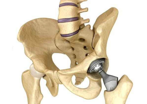
Endoprothetic of the hip jointsIt is used in serious cases of stadium III, when pain syndrome cannot be eliminated and the mobility of the joint is significantly limited.The hip joint prostheses lead to a decrease in pain syndrome, an improvement in the functional state of the joint and the quality of life of the patient.The effect persists for 10-15 years, after which a second operation could be requested.During the surgery, the hip joint is replaced by artificial imitation of ceramics, metal (more often used in titanium) or polymer.
Forecast.Prevention
The prognosis of arthrosis of the hip joints in relation to the patient's life is favorable, but the disease often leads to disabilities.According to the World Health Organization, 80 % of elderly patients with coxarthrosis have a violation of mobility and 25 % cannot do daily issues.In this regard, the primary prevention of arthrosis of the hip joints is important.
Prevention measures:
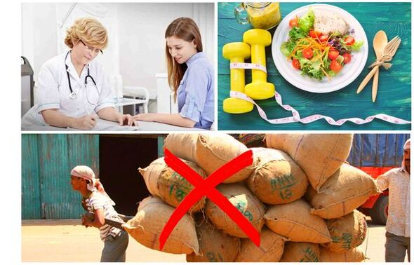
- Reduce body weight.Nutrition must be adjusted to reduce weight and load on the joint.In addition, a decrease in the volume of the adipose tissue reduces the amount of inflammation mediators it has released.
- Avoid heavy physical work and sports overloads.Physical overloads are often the cause of the arthrosis of the hip joints, while moderate physical activity, on the contrary, improves the condition of the joint cartilage, maintains its normal mobility and reduces the load on other joints.
- Correct the disease below.If the patient is detected in diseases that can lead to secondary coksartrosis (endocrine, rheumatic and others), the disease below is necessary.The normalization of the hormonal background and the achievement of the persistent remission of rheumatic diseases is both the primary prevention of arthrosis and allows you to slow down its development.
- Lead a healthy lifestyle.A balanced diet with sufficient content of vegetable and animal proteins, polyunsaturated fatty acids and limiting simple carbohydrates, as well as moderate physical activity, avoiding the occurrence of coaxarthrosis even in the presence of risk factors.
Currently, the prevention of hip joint diseases is mandatory in neonatology and pediatrics.Over time, adequate congenital dysplasia of the hip joint significantly reduces the risk of coxartrosis in adulthood.